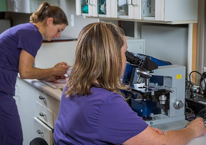
When you can’t visually see the cause of your pet’s pain or discomfort, diagnostic imaging tools are a great resource. Ultrasound and X-rays provide an opportunity to understand what is happening to your pet and determine the best treatment plan. Garibaldi Veterinary Hospital makes use of current medical diagnostic tools such as ultrasound, x-rays, and dental X-rays to diagnose and treat your pet’s health concerns.


Diagnostic lab testing
In-house blood analyzers allow us to quickly obtain diagnostic test results. We can then promptly commence appropriate therapy and treatment. We also conduct urine analysis in-house, which provides us with more accurate and timely results. With these benefits, we can perform routine or emergency surgery knowing your pet’s organs are functioning properly and no hidden health conditions exist that could put your pet at risk while under a general anesthetic. Detecting any abnormalities pre-surgery allows us to alter the anesthetic procedure or take other precautions to safeguard your pet’s health. In addition to our in-house diagnostic equipment, we also work closely with an external laboratory and pathologist for more specialized diagnostic tests in Squamish.
Hip dysplasia
Canine hip dysplasia (abnormal development of the hip joint) begins when the hip joint in a young dog becomes loose or unstable. If left undiagnosed and untreated, this instability causes abnormal wear of the hip cartilage and ultimately progresses to osteoarthritis or degenerative joint disease. Signs of this condition are pain, reluctance to get up or exercise, difficulty climbing stairs, a “bunny-hopping” gait, limping, and lameness, especially after periods of inactivity or exercise.
Hip dysplasia most commonly affects large- and giant-breed dogs; however, smaller dogs can also be affected. Although genetics often play a role in this disorder, young dogs that grow or gain weight too quickly or get too much high-impact exercise are also at risk. Being overweight can aggravate hip dysplasia.
We can help prevent or slow this condition by monitoring food intake and ensuring that your dog gets proper exercise as he or she ages. We can also screen your dog for hip dysplasia, using one of two methods. The earlier we can diagnose hip dysplasia, the better the possible outcome for your dog.
OFA (Orthopedic Foundation for Animals) Certification:
We can x-ray your dog’s hips for hip dysplasia at 2 years of age. We will forward these radiographs to the OFA, where board-certified radiologists will evaluate and grade your dog’s hips for OFA certification. Correct positioning of your dog is essential for proper radiographic evaluation, so a general anesthetic is required to make the procedure less stressful for him or her.
PennHIP Method:
We can x-ray your dog’s hips using the PennHIP method for evaluating hip dysplasia in dogs, which can be performed much earlier (at 16 weeks of age) than OFA certification. Requiring a general anesthetic, it involves x-raying your dog’s hips in three different positions to measure how loose the joints are and determine the presence or likelihood of osteoarthritis. If you are a breeder, consider using this test to help you select good breeding candidates at a younger age. If your dog competes athletically, consider using this technique to evaluate the future soundness of your dogs or puppies.
Please call us to discuss your dog’s risk of developing hip dysplasia, to schedule a screening, or to discuss treatment options.
Radiography (x-Ray)
We are pleased to offer the quality and efficiency of digital radiographs. The images are obtained through the use of a digital plate, which then converts the X-ray into an image that can be viewed on a computer screen. The images can then be modified to increase diagnostic value and can also be emailed for further consultation. Digital X-rays are a very useful tool that can be used to evaluate bones, joints, soft tissue structures, and abdominal and chest cavities.
Ultrasound
Ultrasound is an important diagnostic tool that allows us to visualize soft tissue structures (such as abdominal organs) in an extremely detailed, yet non-invasive and pain-free manner.
For more information, please read our What to Expect Pet Ultrasound document.
What to Expect Pet Ultrasound